My screening pathway
Breast cancer screening, step by step
Here is how organised screening works in France, from the invitation to the results. Each point links to its official source.
English translation of official French information. The original source takes precedence.
Step 1
Your invitation
Taking part is a personal choice.
 Ask a question about this step
What you need to know
Your invitation arrives by post and is also available in your ameli account. It is kept there for 6 months, then automatically deleted.
AmeliUpdated on May 11, 2026
All the information and sources
Between the ages of 50 and 74, you are invited for a screening mammogram every 2 years. Taking part is a personal choice.
Organised screening is for women aged 50 to 74 who have no symptoms and no particular risk factor for breast cancer other than their age.
AmeliUpdated on May 11, 2026
Taking part in screening is a personal choice. Read about its benefits and drawbacks before deciding.
AmeliUpdated on May 11, 2026
If you are 50 and have not received your invitation, or if you have lost it, you can find it in your ameli account or ask your usual doctor (médecin traitant) for a new invitation.
AmeliUpdated on May 11, 2026
Frequently asked questions at this stage
Step 2
Preparing for your appointment
Choose a radiologist from the list of approved radiologists included with your invitation and make an appointment.
 Ask a question about this step
What you need to know
Bring your carte Vitale and your screening invitation letter to the appointment.
AmeliUpdated on May 11, 2026
All the information and sources
Choose a radiologist from the list of approved radiologists included with your invitation and make an appointment.
Choose your radiologist from the list included with the invitation letter. You can check whether your usual practice is on the list.
AmeliUpdated on May 11, 2026
You have nothing to pay for the mammogram under the organised screening programme. Assurance Maladie pays the radiologist directly.
AmeliUpdated on May 11, 2026
Bring all the images and reports from previous breast examinations. They allow the doctor to compare the images and information.
AmeliUpdated on February 26, 2026
If you have not reached menopause, arrange the examination within the fifteen days following the start of your period. Your breasts are less sensitive during this time.
AmeliUpdated on February 26, 2026
On the day of the appointment, do not apply cream, powder, perfume or deodorant to your underarms or breasts. These products can interfere with the examination and the reading of the results.
AmeliUpdated on February 26, 2026
Frequently asked questions at this stage
Step 3
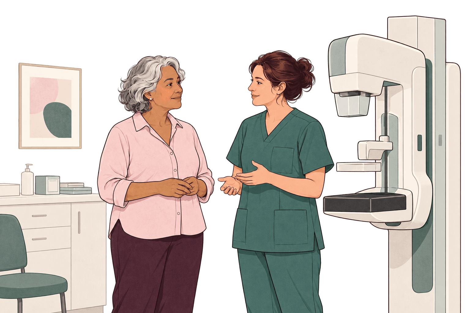
During the mammogram
The radiologist also examines your breasts by looking at and feeling them.
 Ask a question about this step
What you need to know
If you are worried about the examination or have had a painful experience before, speak to the radiologist or radiographer.
INCaUpdated on September 26, 2025
All the information and sources
A mammogram is an X-ray examination of the breasts that uses a low dose of radiation. The radiologist also examines your breasts by looking at and feeling them.
The examination itself takes about fifteen minutes.
AmeliUpdated on February 26, 2026
Each breast is placed between 2 plates that compress it for a few seconds. The radiographer takes 2 images of each breast.
INCaUpdated on September 26, 2025
The radiologist looks at and feels your breasts: this is the clinical examination. They look for abnormalities that are sometimes not visible on the mammogram.
INCaUpdated on September 26, 2025
If you are pregnant or might be pregnant, tell the radiologist. Special precautions are needed to protect the fetus from X-rays.
AmeliUpdated on February 26, 2026
Frequently asked questions at this stage
Step 4
Reading the images
The radiologist analyses your images.
 Ask a question about this step
What you need to know
Mammograms considered normal are systematically reviewed by a second expert radiologist as part of organised screening.
AmeliUpdated on May 11, 2026
All the information and sources
The radiologist analyses your images. In organised screening, mammograms considered normal are reviewed by a second expert radiologist.
After the mammogram, the radiologist analyses your images, meaning the pictures taken during the examination.
AmeliUpdated on February 26, 2026
The radiologist may add further images or an ultrasound to the mammogram to help interpret it. This does not necessarily mean there is an abnormality.
INCaUpdated on September 26, 2025
Frequently asked questions at this stage
Step 5
Receiving your results
The radiologist gives you the initial results at the end of the consultation.
 Ask a question about this step
What you need to know
You receive the final results by post. They are also sent to the doctor of your choice.
AmeliUpdated on May 11, 2026
All the information and sources
The radiologist gives you the initial results at the end of the consultation. The final results are sent by post to you and to the doctor of your choice.
The radiologist gives you the initial results at the end of your consultation.
AmeliUpdated on May 11, 2026
The final results are available from your radiologist within 2 weeks.
INCaUpdated on September 26, 2025
If abnormalities are identified on the images, further examinations may be requested, such as an ultrasound.
AmeliUpdated on May 11, 2026
Additional examinations are reimbursed by Assurance Maladie and your complementary health insurer (mutuelle) under the usual conditions, excluding any fees charged above the standard rate.
AmeliUpdated on May 11, 2026
Keep the images and report from each mammogram. Bring your previous mammograms to future screening appointments so the radiologist can compare them.
AmeliUpdated on May 11, 2026
Frequently asked questions at this stage
Step 6
Between screening appointments
Between mammograms, remain alert to changes in your breasts.
 Ask a question about this step
What you need to know
Have your breasts examined at least once a year by your usual doctor (médecin traitant), midwife or gynaecologist.
AmeliUpdated on May 11, 2026
All the information and sources
If no abnormality is detected at the first or second reading, you will be invited to repeat the examination in 2 years. Between mammograms, remain alert to changes in your breasts.
If no abnormality is detected at the first or second reading, you will be invited to repeat the examination in 2 years.
INCaUpdated on September 26, 2025
Between mammograms, consult a healthcare professional if you notice a lump or swelling in a breast or an armpit.
AmeliUpdated on May 11, 2026
Consult a healthcare professional if you notice redness or an uneven appearance of the skin on your breasts, such as an “orange peel” texture.
AmeliUpdated on May 11, 2026
If you notice that your nipple is pulling inwards, consult a healthcare professional.
AmeliUpdated on May 11, 2026
Consult a healthcare professional if you notice abnormal discharge.
AmeliUpdated on May 11, 2026
Frequently asked questions at this stage
Have a question about a stage?
Describe your question in your own words. AskPink answers using the same sources.
Ask a questionThis pathway describes organised screening in France. Your situation may be different: your doctor remains your best point of contact.